FLIM experiments from start to finish
- Image acquisition
- Data analysis
- Hardware control & automation
SPEED
unprecedented frame rates up to 300 fps with FLIM images at 100 fps
SENSITIVITY
excellent light sensitivity with advanced fluorescence lifetime imaging capabilities
SIMPLICITY
automatic data analysis instantly calculates the fluorescence lifetime
Fluorescence Lifetime Imaging Microscopy (FLIM) has become an important tool to assess the biochemical environment of fluorescent molecules and probes.
Upon excitation, fluorescent molecules emit light and the fluorescent lifetime quantifies the decay rate of that emitted light. The fluorescence lifetime is a telltale signature of the molecules and their immediate environment.
FLIM is the technique which maps the spatial distribution of lifetimes in living cells and in inorganic material. Fluorescence lifetime is independent of concentration, bleaching and intensity variations, making it an inherently quantitive technique, and a key advantage over the light intensity.
Applications
-
- Molecular interactions
- FRET/FLIM imaging
- Biosensors
- NADH/FAD fluorescence dynamics
- Membrane dynamics
- Viscosity imaging
- LED inspection
- Solar cell MCL monitoring
- ...and many more!

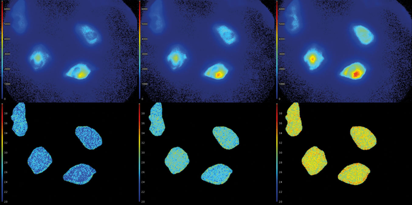
Top Row
Light intensitty images (colourised).
Bottom Row
Corresponding fluorescence lifetime images (colourised). The average fluorescence lifetime of the cells increases over time.
Image courtesy of the Netherlands Cancer Institute

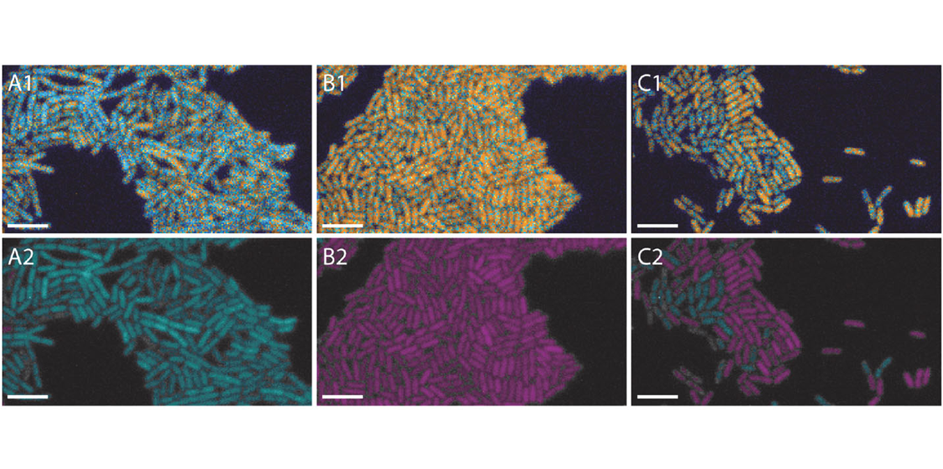
B.subtilis cells showing different lifetimes.
Top Row
Original lifetime image of FRETing, non-FRETing, and mixed cells.
Bottom Row
Same cells as top row, but categorised and colorised based on avarage cell life-time. Scale vbar is 5um.
Image courtesy of University of Groningen

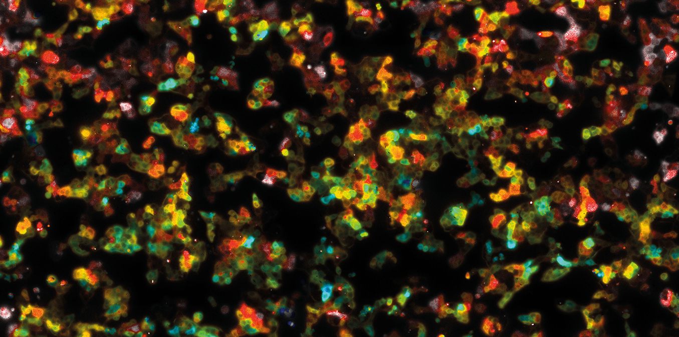
Stitched lifetime image of Hap 1 cells expressing EPAC-sensor that shows a lifetime increase when cAMP concentration increases after stimulation with isoproternol.
Image courtesy of the Netherlands Cancer Institude
LIFA FLIM SYSTEM
A camera-based FLIM system for fast fluorescence lifetime imaging.
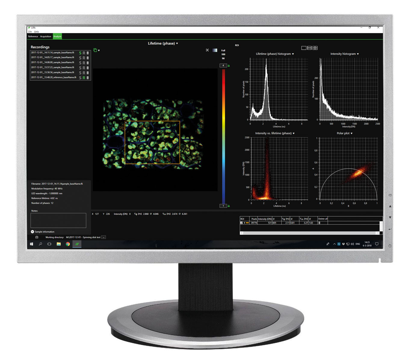
With the Lambert Instruments FLIM Attachment (LIFA), you record quantitive lifetime data in a matter of seconds. It’s easy to connect our FLIM camera and light source to your microscope, and our specialised software records the images and analyses the data instantly, presenting the results visually for easy interpretation.
LEARN MORE
User Publications
Researchers around the world use the LIFA in their studies. Opposite is an overview of the most recent publications describing research that was done using a LIFA.
For a complete overview of applications, please visit our applications pages and our selected LIFA publications on the Lambert main site for more information.
Mutant APC reshapes Wnt signaling plasma membrane nanodomains by altering cholesterol levels via oncogenic β-catenin
July 19 - FLIM
Electrocatalytic Reaction Driven Flow: Role of pH in Flow Reversal
Mar 2, 2022 - FLIM, proton concentration, University of Twente
Bioorthogonal labeling of transmembrane proteins with non-canonical amino acids unveils masked epitopes in live neurons
Dec 7, 2021 - Microscopy, Fluorescence
Quantification of PD-1/PD-L1 Interaction between Membranes from PBMCs and Melanoma Samples Using Cell Membrane Microarray and Time-Resolved Förster Resonance Energy Transfer
Dec 7, 2021 - FRET, Fluorescence
Glycerol suppresses glucose consumption in trypanosomes through metabolic contest
Aug 13, 2021 - FLIM
PIM kinases inhibit AMPK activation and promote tumorigenicity by phosphorylating LKB1
Jun 30, 2021 - FLIM